Highly effective even in the low oxygen environment of tumors.
Radiation kills tumors by creating oxygen free radicals that damage the tumor’s DNA. However, the lack of oxygen in the center of tumors blocks the production of free radicals, inhibiting destruction by radiation. NIBIB researchers have now designed a nanoparticle that generates radiation-induced oxygen free radicals even in the core of low-oxygen tumors, dramatically increasing the success of radiotherapy.
Low-oxygen (hypoxic) regions in the core of tumors produce strong resistance to radiation therapy, leading to cancer invasion and metastasis. Hypoxia, a common feature of solid tumors, occurs in the central region of most cancers, so developing therapies that overcome the problem of tumor hypoxia is a critical goal to increase the effectiveness of cancer therapy.
At the NIBIB Laboratory of Molecular Imaging and Nanomedicine (LOMIN), led by Xiaoyuan (Shawn) Chen, Ph.D., principal investigator, one of the main areas of research is the creation of nanoparticles that can target and kill tumors without harming surrounding healthy tissues. To that end, the lab has created several nanoparticles, many of which contain chemotherapy drugs such as doxorubicin and deliver them to the tumor site.
In the current study led by Wenpei Fan, Ph.D., a postdoctoral fellow in the LOMIN laboratory, the nanoparticle carries a different class of molecules: compounds designed to work with radiation and enhance its ability to kill tumor cells. The work appears in the March issue of the journal Nature Communications.1
The nanoparticle they designed is called hollow mesoporous organosilica nanoparticle (HMON). Mesoporous refers to the size of the nanoparticle’s pores, through which anticancer compounds can be loaded and then released when they reach the tumor. A mesoporous material contains pores with a diameter between 2 and 50 nanometers. For comparison, microporous materials have pores smaller than 2 nanometers.
Organosilica refers to the basic structure, which is silica, a material like quartz. Specific organic molecules are added to the organosilica mixture to create nanoparticles with the desired pore size to load the anti-cancer compounds.
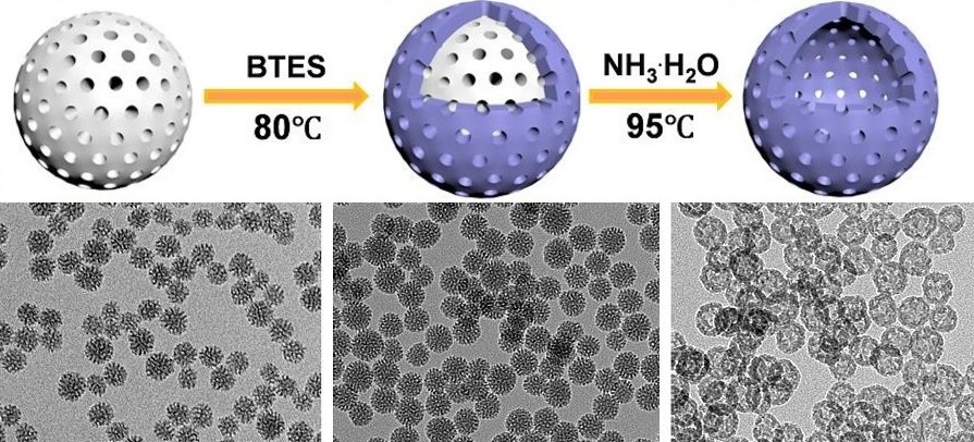
The researchers used a special technique called ammonia-assisted hot water etching to design very small nanoparticles. Creating very small nanoparticles was important because smaller particles could travel to the center of the tumor, which was the main goal of these experiments.
The small particles were made by starting with a very small central bead that was basically the mold for the nanoparticle layer that was attached to the outside of the bead. However, nanoparticles must be hollow to carry their cancer-killing payload. This is where ammonia-assisted hot water etching came into play. This particular method was used because it removes or degrades the center bead but does not damage the outer coating. In this way, the central bead was removed leaving the hollow mesoporous nanoparticle very small.
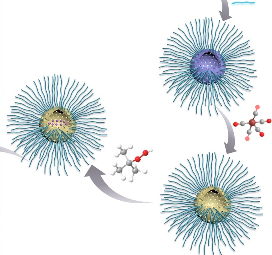
The nanoparticles were then loaded with two different compounds. A compound creates oxygen free radicals in the oxygen-rich environment of a tumor when it receives radiation, which are the outer portions of the tumor. The other compound is capable of creating oxygen free radicals when irradiated in the hypoxic core of the tumor. Oxygen free radicals are very reactive and damage the DNA of tumor cells, killing the cancer cells.
The loaded nanoparticles were tested on a human glioblastoma cell line to determine the amount of DNA damage generated when combined with radiation. A test that stains DNA fluorescent red clearly showed that the combination of nanoparticles and radiation destroyed the DNA of glioblastoma cells compared to nanoparticles alone or radiation alone. Additional testing showed that the nanoparticles generated oxygen free radicals when exposed to radiation under both normal and very low oxygen conditions, confirming that the system worked as designed.
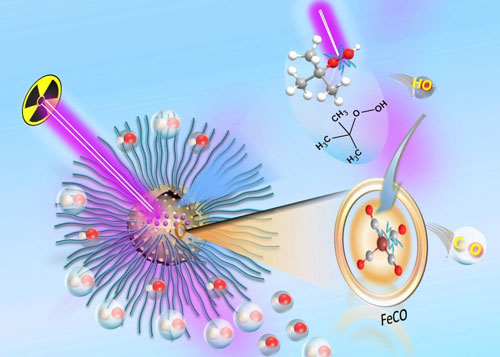
The destruction of cancer cells by HMONs was further aided by the fact that the two compounds loaded into the nanoparticles react with each other with radiation. The result is the production of carbon monoxide gas, which is toxic to cancer cells. The combination of scavenging oxygen free radicals and removing carbon monoxide using radiation resulted in what researchers called radiodynamic therapy or gas therapy/RDT.
Gas and PDR therapy was tested on mouse tumors. When the nanoparticle was injected into the bloodstream of mice without radiation, the effect on the tumors was minimal. Radiation alone also had a minimal effect. However, when the nanoparticle was injected and then activated by radiation, the tumors shrank dramatically without toxic effects on the mouse organs.
Lead author Fan says, “We believe this gas therapy/RDT approach offers new possibilities for enhanced X-ray-activated treatment for future deep cancer therapies. The next logical step is to optimize the structure and scale up nanoparticle synthesis to enable clinical translation of this type of enhanced radiotherapy.”
The project was led by the Laboratory of Molecular Imaging and Nanomedicine, National Institute of Biomedical Imaging and Bioengineering (NIBIB), National Institutes of Health, Bethesda, MD and the Laboratory of Cellular Imaging and Macromolecular Biophysics, NIBIB, Bethesda, MD in cooperation with collaborators from the Department of Radiology, the Second Affiliated Hospital, Zhejiang University School of Medicine, Zhejiang, China, the Department of Medical Imaging, Jinling Hospital, Nanjing University College of Medicine, Jiangsu, China, the Institute of Radiological Medicine, Fudan University, Shanghai, China, the State Key Laboratory of High Performance Ceramics and Superfine Microstructure, Shanghai Institute of Ceramics, Chinese Academy of Sciences, Shanghai, China, and the Laboratory of Intelligent Hybrid Materials, Center for Advanced Membranes and Porous Materials, King Abdullah University of Science and Technology, Thuwal, Saudi Arabia.