
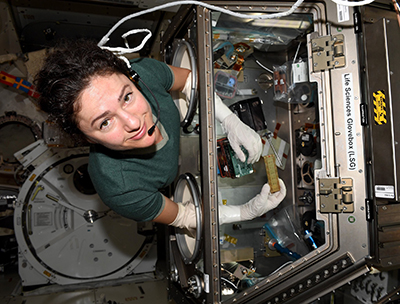
Shortly after 3:30 pm EDT on April 7, 2020, the SpaceX Dragon spacecraft splashed down in the Pacific Ocean and returned to Earth after nearly a month docked with the International Space Station (ISS). The spacecraft brought back a number of physical and life sciences research projects, including heart and intestinal tissue chip projects that traveled to the ISS as part of the Tissue Chips in Space initiative.
The initiative is an ambitious collaborative effort bringing together the National Center for Advancing Translational Sciences (NCATS), the National Institute of Biomedical Imaging and Bioengineering (NIBIB), and the ISS US National Laboratory to rapidly advance tissue chip technology for biomedical research.
The chips contain miniature models of living tissues built from human cells. Because tissue chips accurately model the structure and function of human organs and systems, researchers could use them to predict (more quickly and effectively than current methods allow) whether a candidate drug, vaccine or biological agent is safe or toxic in humans. Tissue chips may also provide new opportunities for researchers to study biological processes in the extreme microgravity environment aboard the ISS.
Prolonged exposure to microgravity, such as that experienced by astronauts on the ISS, is known to cause changes in many aspects of the human body’s functioning, including the heart and cardiovascular system. Many of the changes caused by microgravity appear similar to those caused by accelerated diseases and aging processes. The use of tissue chips on the ISS provides a unique opportunity for researchers to model and study conditions related to disease and aging over weeks or months, rather than the years it would take for these conditions to develop on Earth.
A research team led by Deok-Ho Kim, Ph.D., of Johns Hopkins University and comprised of researchers from several academic institutions and other organizations, developed heart tissue chips to study structural and functional changes in tissue caused by spending time in low gravity. The team also developed the technology needed to safely send the tissue chips into space, survive aboard the ISS for nearly a month, and return to Earth. The researchers hypothesized that during that period of time tissues would undergo functional, visible, and molecular changes similar to those that occur with disease or old age on Earth.
To create the heart tissue chips, the researchers convinced human-induced pluripotent stem cells, which have the potential to become almost any type of cell in the body, to form cardiomyocytes (heart muscle cells) that grew into small three-dimensional structures between two posts on a silicon scaffold. One of the poles, which was flexible and flexed every time the tissue contracted, contained a small magnet. While on the ISS, the magnet’s movements were captured by a sensor and transmitted to researchers on the ground so they could collect data on the strength of the contractions (and any changes in that strength) remotely.
Now that the tissue chips have returned to Earth, researchers will continue to analyze the data they collected while the tissue chips were on the ISS and compare it to data from a set of comparison chips that remained on Earth. The research team will also begin to evaluate visible and molecular changes that the tissue may have experienced as a result of spending time in microgravity.
In a future flight, researchers will test methods to improve heart cell contractions, including mechanical stimulation, which mimics muscle conditioning, as well as drugs known to protect the heart from irregular heartbeats or heart disease. The project’s findings may have implications both for improving the health of astronauts and for finding new ways to study and treat heart diseases related to aging on Earth.
For more photos, go to NCATS coverage. here.
This work was funded with the support of NIBIB and NCATS (UG3 EB028094).


